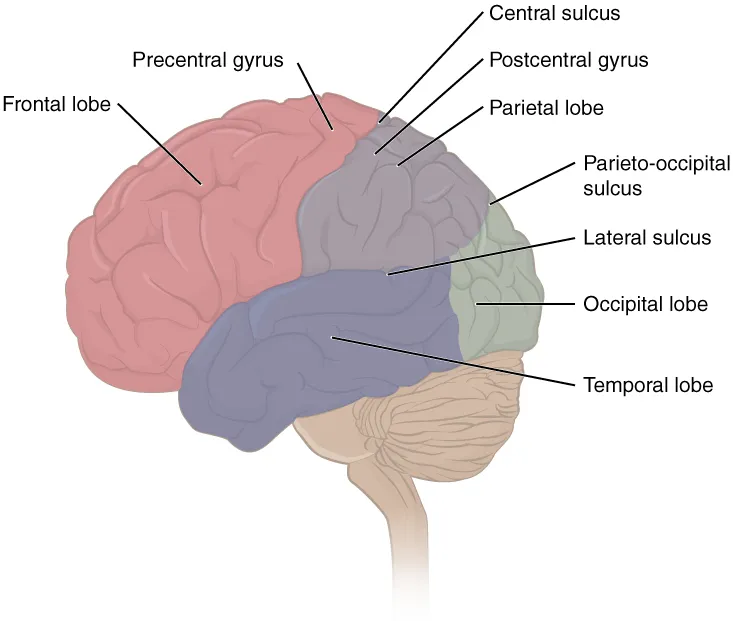
Head & Neck — Layer-by-Layer Anatomy
Explore the anatomical planes from the external surface to the cranial cavity. Select a layer to study its components, relationships and clinical significance.
Layer explorer
Essential anatomical orientation
- Anterior: toward the front; posterior: toward the back.
- Superior / inferior: above / below.
- Medial / lateral: toward / away from the midline.
- Superficial / deep: nearer to / farther from the body surface.
- Ipsilateral / contralateral: same side / opposite side.
Study method: identify the layer, name the structure, describe its function, then trace its arterial supply, venous drainage and nerve supply.
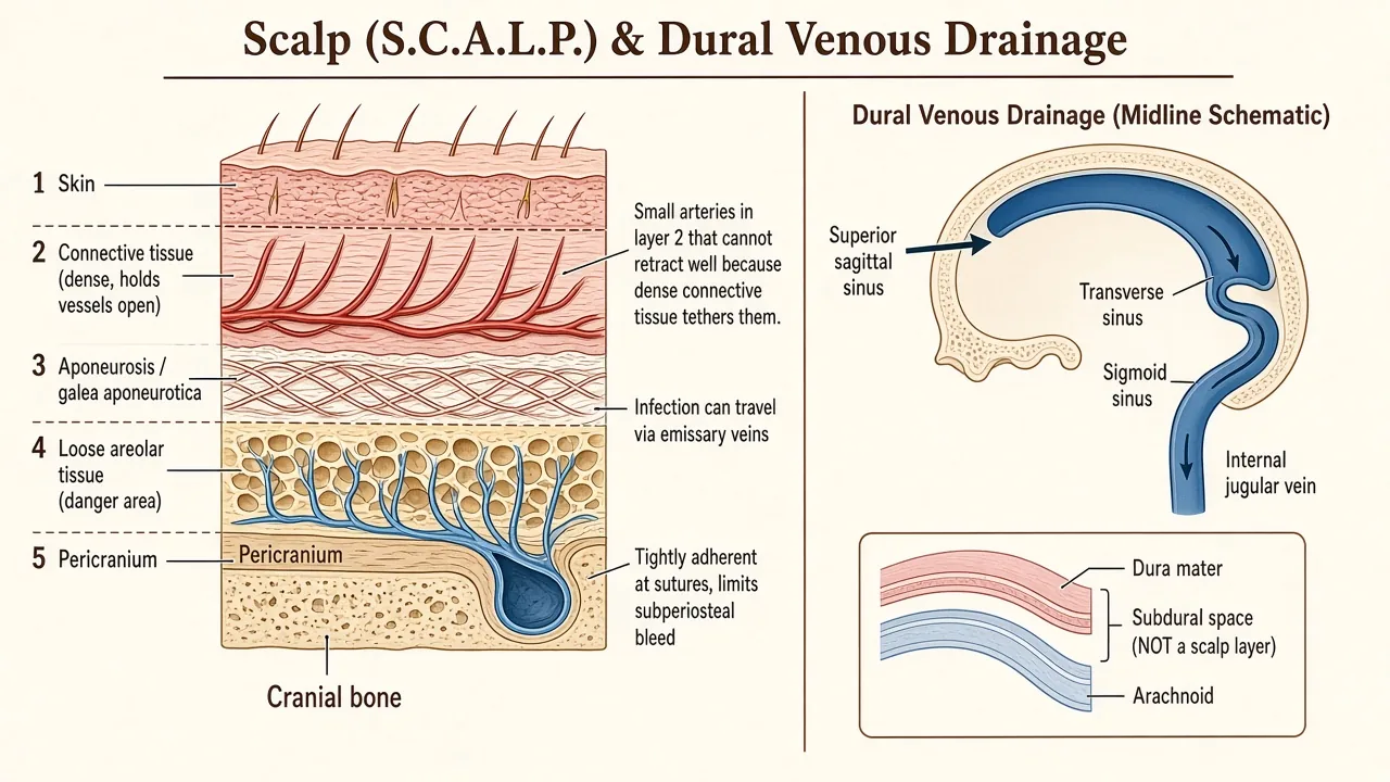
Scalp: the five anatomical layers — S.C.A.L.P.
| Layer | Medical term | Key features | Clinical relevance |
|---|---|---|---|
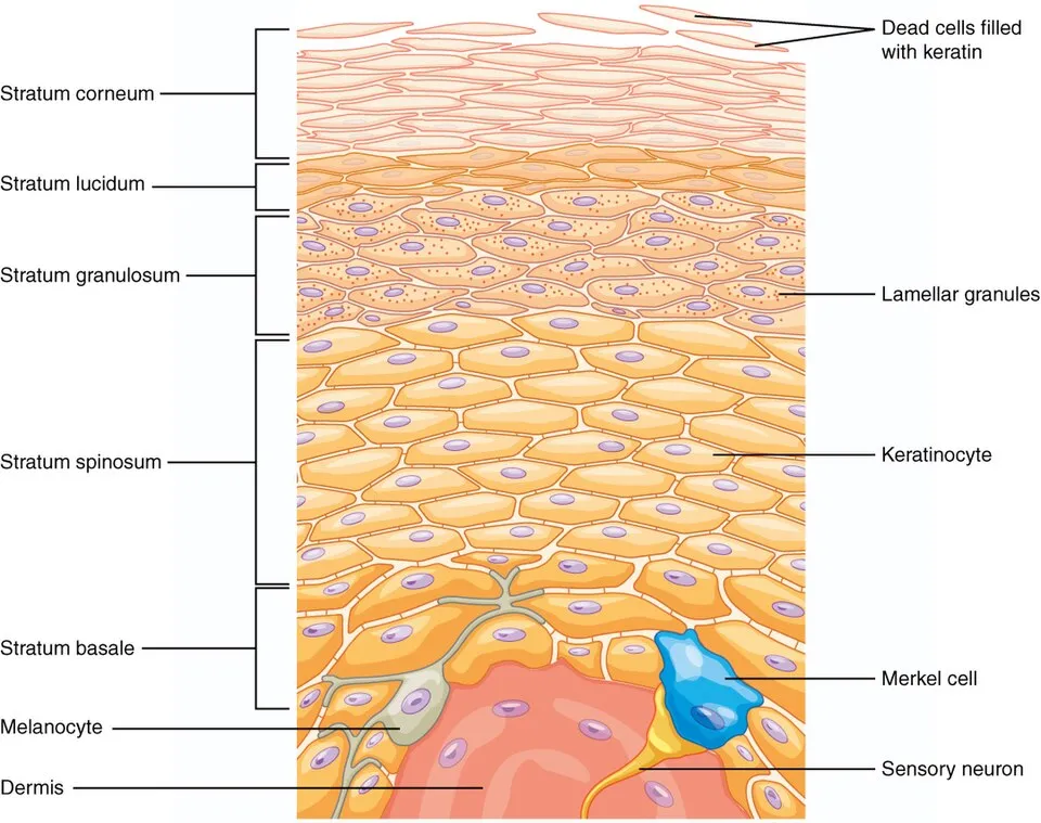
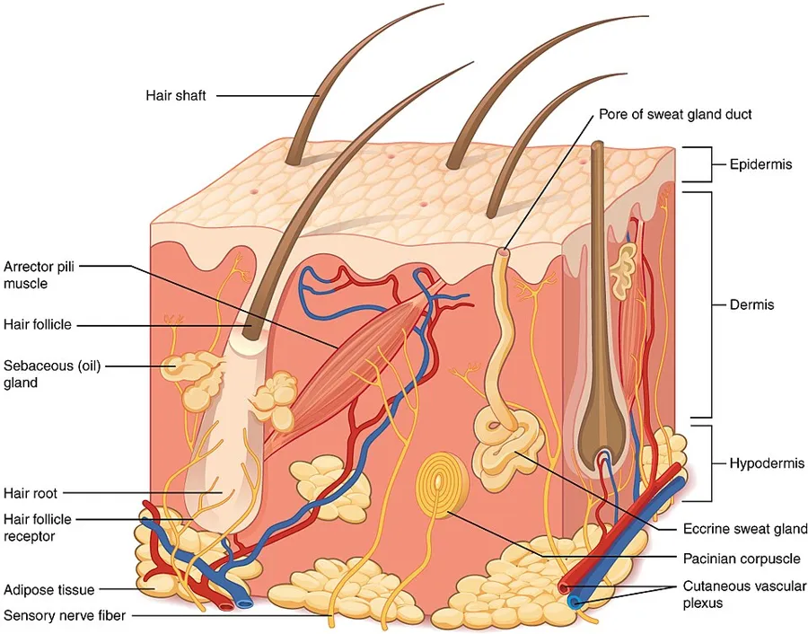
| S | Skin | Hair follicles, sebaceous glands, sweat glands; dense hair-bearing skin. | Scalp lacerations may bleed profusely because vessels are held open by dense connective tissue. |
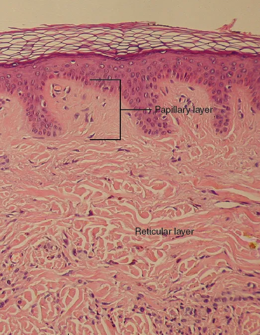
| C | Connective tissue | Dense, fibrofatty superficial fascia containing cutaneous nerves and vessels. | Fibrous septa tether vessels, limiting vasoconstrictive retraction after injury. |
| A | Aponeurosis — galea aponeurotica | Tendinous sheet connecting frontalis and occipitalis muscles; part of the epicranius. | Full-thickness wounds crossing the galea may gape due to muscle pull. |
| L | Loose areolar connective tissue | Mobile plane between galea and pericranium; contains emissary veins. | Potential “danger area” because infection can spread via emissary veins to intracranial venous sinuses. |
| P | Pericranium | Periosteum covering the external surface of cranial bones. | Subperiosteal bleeding is limited by suture lines where periosteum is firmly attached. |
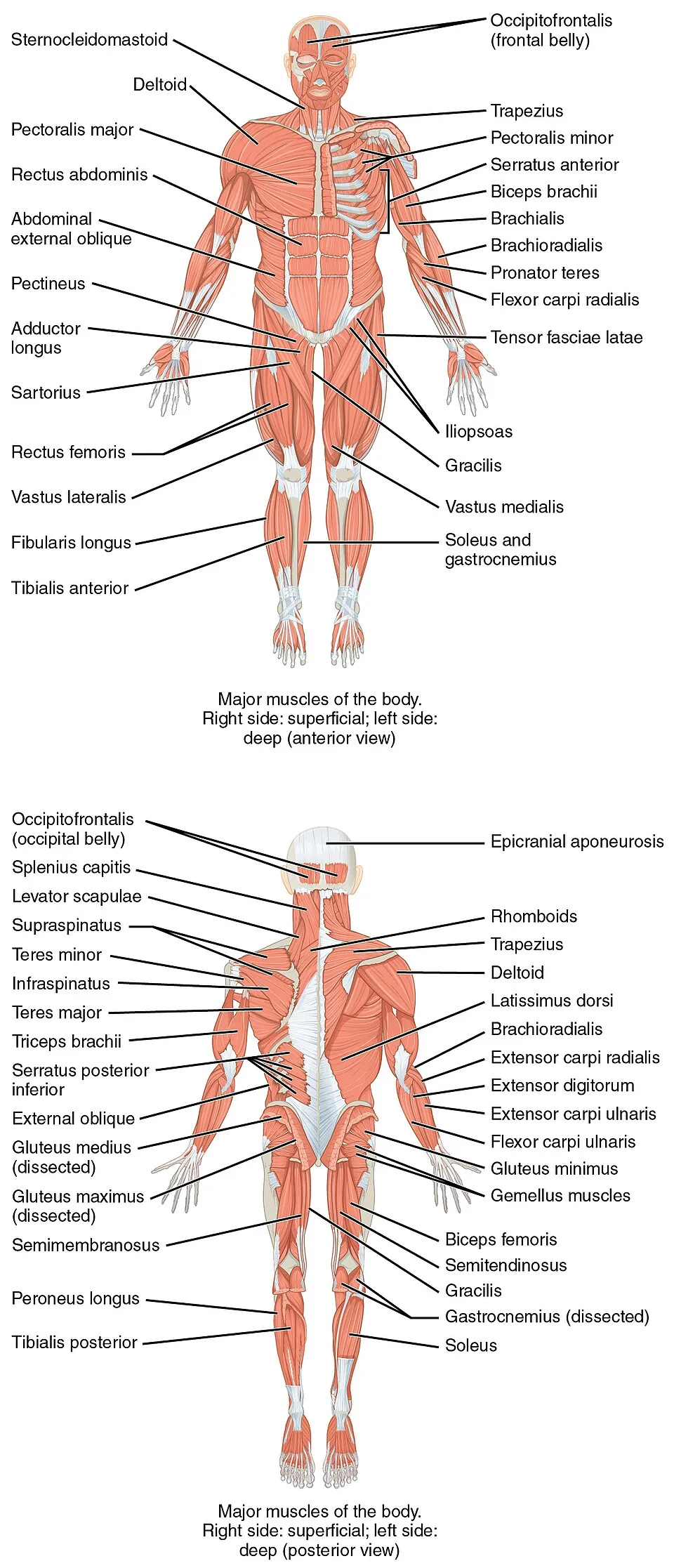
Facial muscles — muscles of expression
Innervation: Facial nerve (CN VII).
- Orbicularis oculi: closes eyelids; palpebral and orbital parts.
- Orbicularis oris: closes and protrudes lips.
- Buccinator: compresses cheek; keeps food between occlusal surfaces.
- Zygomaticus major/minor: elevates upper lip and mouth angle.
- Frontalis: elevates eyebrows and wrinkles forehead.
- Platysma: tenses skin of anterior neck and depresses mandible/lower lip.
Muscles of mastication
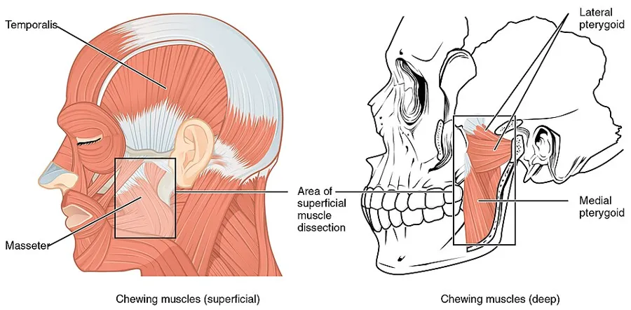
Innervation: Mandibular division of trigeminal nerve (CN V3).
- Masseter: elevates mandible; powerful jaw closure.
- Temporalis: elevates and retracts mandible.
- Medial pterygoid: elevates and assists protrusion.
- Lateral pterygoid: protrudes and assists depression; contributes to side-to-side movement.
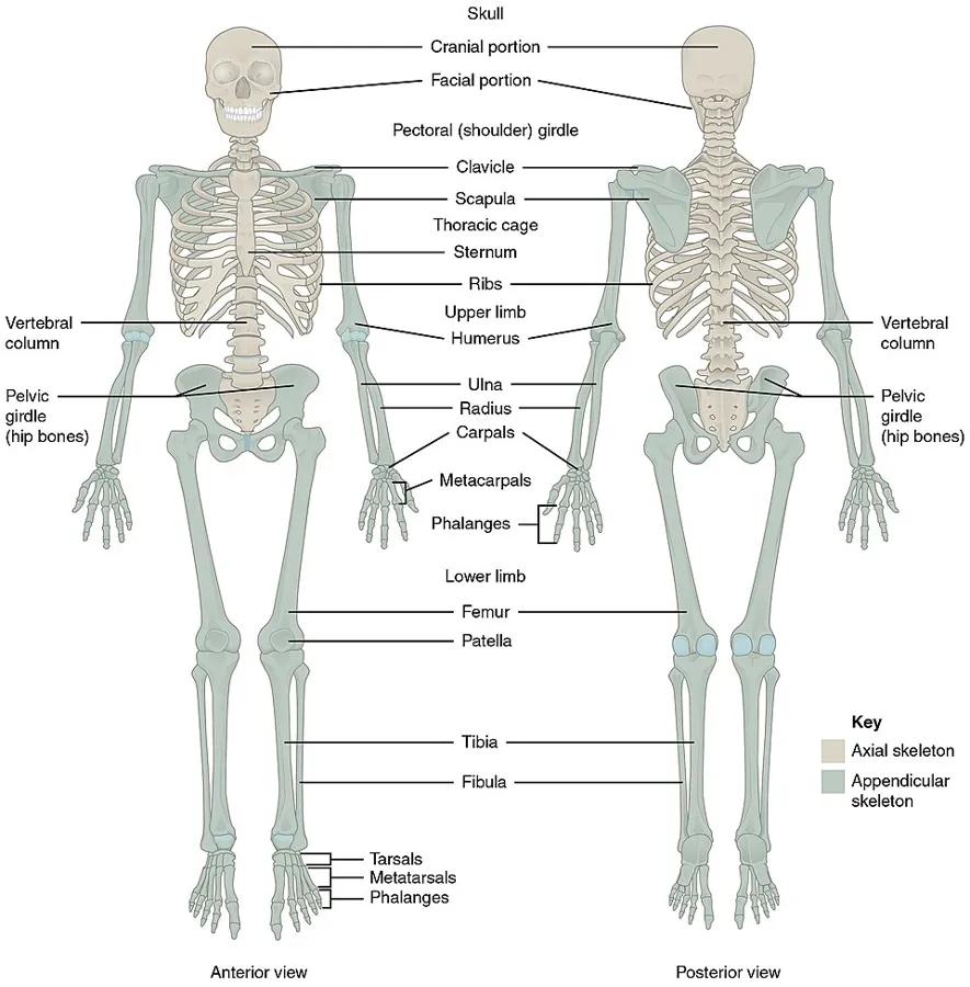
Major cranial bones
- Neurocranium: frontal, parietal (paired), temporal (paired), occipital, sphenoid and ethmoid bones.
- Viscerocranium: maxillae, zygomatic, nasal, lacrimal, palatine, inferior nasal concha, vomer and mandible.
- Landmarks: bregma, lambda, pterion, asterion, supraorbital margin, mastoid process and foramen magnum.
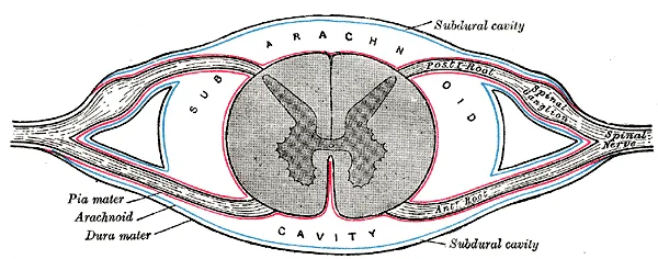
Meninges & intracranial spaces
- Dura mater: periosteal and meningeal layers; forms dural venous sinuses.
- Arachnoid mater: avascular membrane; CSF circulates in the subarachnoid space.
- Pia mater: delicate vascular membrane closely adherent to brain surface.
- Epidural space: potential space in cranium; middle meningeal artery injury may cause epidural haematoma.
- Subdural space: potential space; tearing of bridging veins may cause subdural haematoma.
Neurovascular anatomy of the head
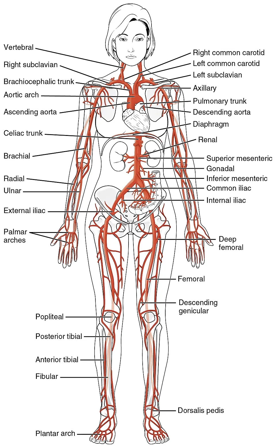
Arterial supply
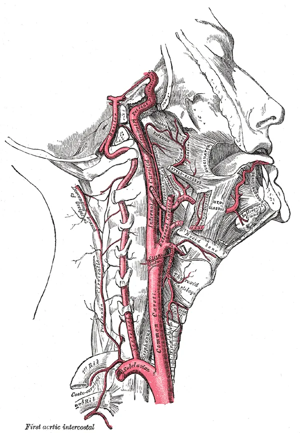
- Internal carotid artery (ICA): contributes to anterior cerebral circulation via anterior cerebral artery (ACA) and middle cerebral artery (MCA).
- Vertebral arteries: unite to form basilar artery; supply posterior circulation through posterior cerebral arteries (PCA) and branches.
- External carotid artery (ECA): supplies much of the face and scalp; branches include superficial temporal, maxillary, facial and occipital arteries.
- Circle of Willis: arterial anastomotic ring at the base of the brain.
Venous drainage
- Dural venous sinuses: superior sagittal, straight, transverse, sigmoid and cavernous sinuses.
- Internal jugular vein (IJV): major venous outflow from cranial cavity.
- Emissary veins: connect extracranial veins with dural venous sinuses.
Cranial nerves — high-yield overview
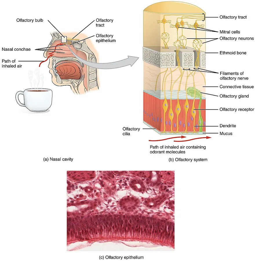
- CN I Olfactory: smell.
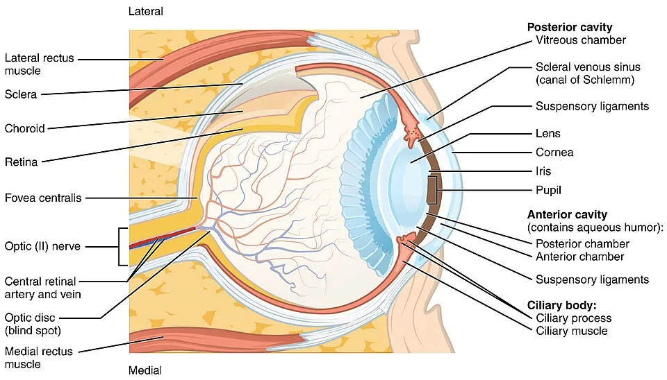
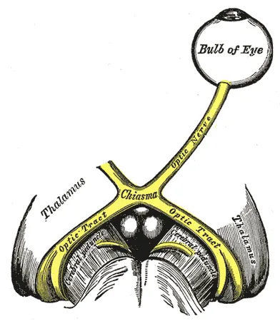
- CN II Optic: vision.
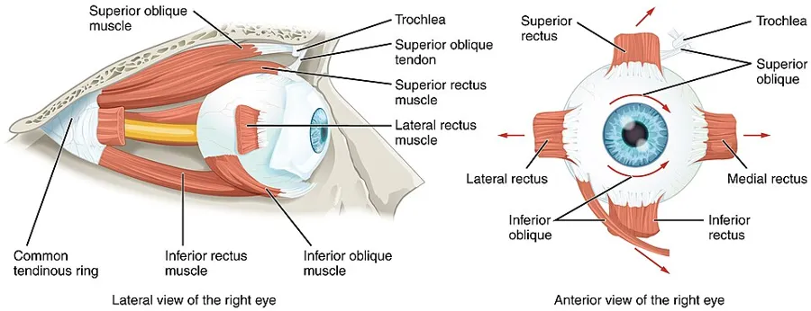
- CN III, IV, VI: most extraocular movements.
- CN V Trigeminal: facial sensation; mastication (V3).
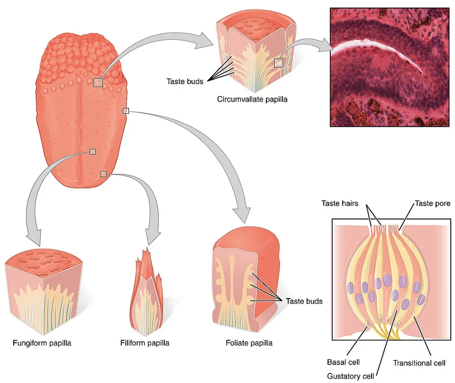
- CN VII Facial: facial expression; taste anterior 2/3 tongue.
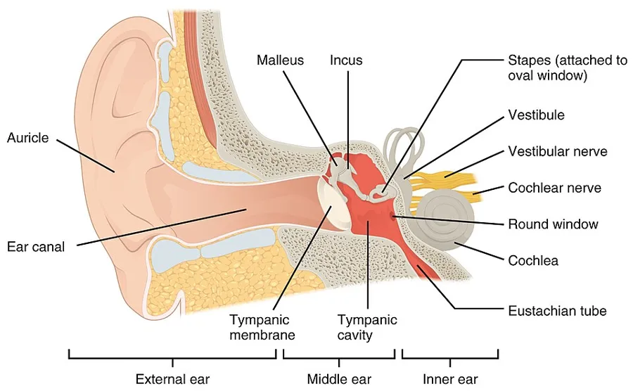
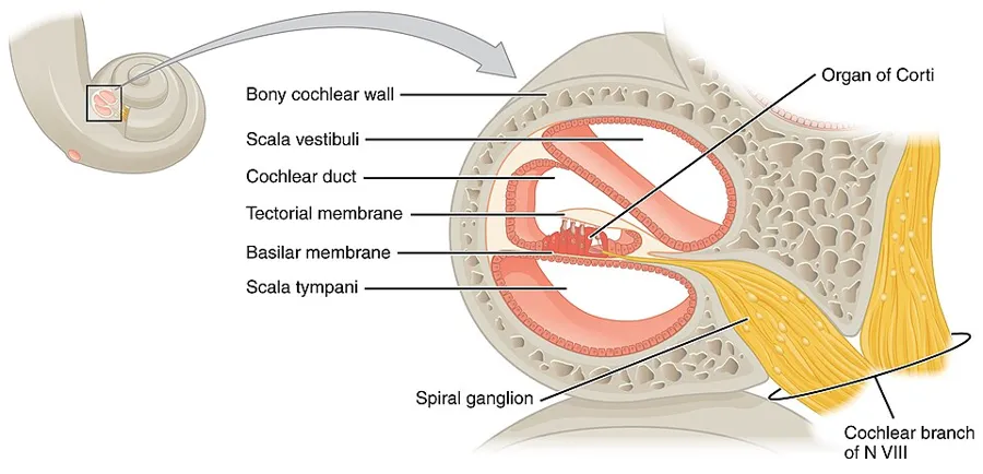
- CN VIII Vestibulocochlear: hearing and balance.
- CN IX, X: swallowing, phonation and visceral functions.
- CN XI Accessory: sternocleidomastoid and trapezius.
- CN XII Hypoglossal: tongue movement.
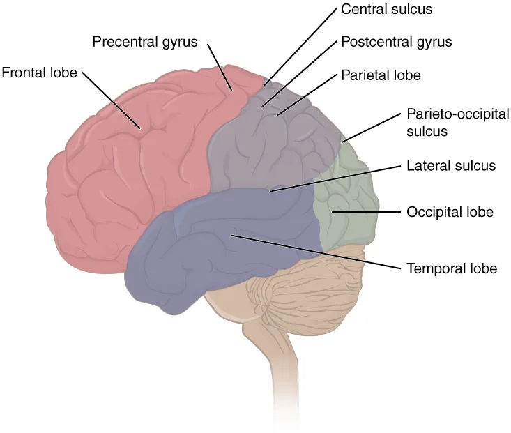
Clinical correlation — stroke: MCA territory infarction commonly produces contralateral face/arm-predominant weakness or sensory loss; dominant-hemisphere involvement may cause aphasia, while non-dominant involvement may cause neglect. Clinical presentation depends on the exact vascular territory and collateral circulation.